
Oodiniasis, or Marine Velvet Disease
White dots, fast breathing and few remedies: the disease that kills the most fish in aquariums
Oodiniasis, or Velvet disease, is the condition that causes the largest number of fish deaths in aquariums.
It should be stressed that there are few remedies for this particular disease.
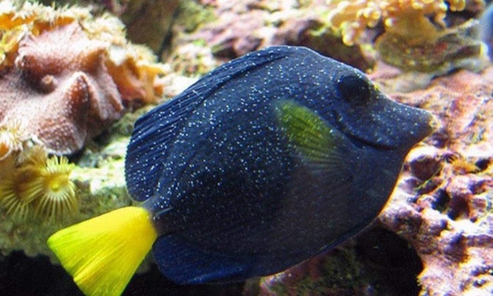
The parasite
In marine fish it is caused by a pathogen belonging to the Dinoflagellate family: Oodinium ocellatum. In freshwater fish the pathogen is instead Oodinium pillularis.
The parasite in question attacks the mucous membranes of infected fish, and so it can attack the skin, the gills and, more rarely, the gut.
Where it attacks the fish
- 1Skin becomes covered in small white dots
- 2Gills breathing speeds up
- 3Gut affected more rarely
The parasite attacks the fish's mucous membranes: skin, gills and, more rarely, gut.
How to recognise it
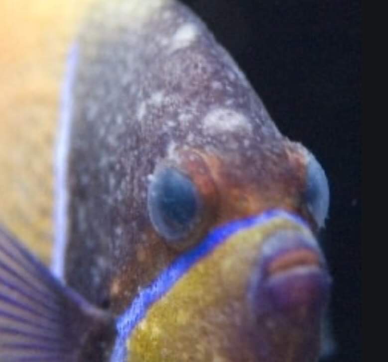
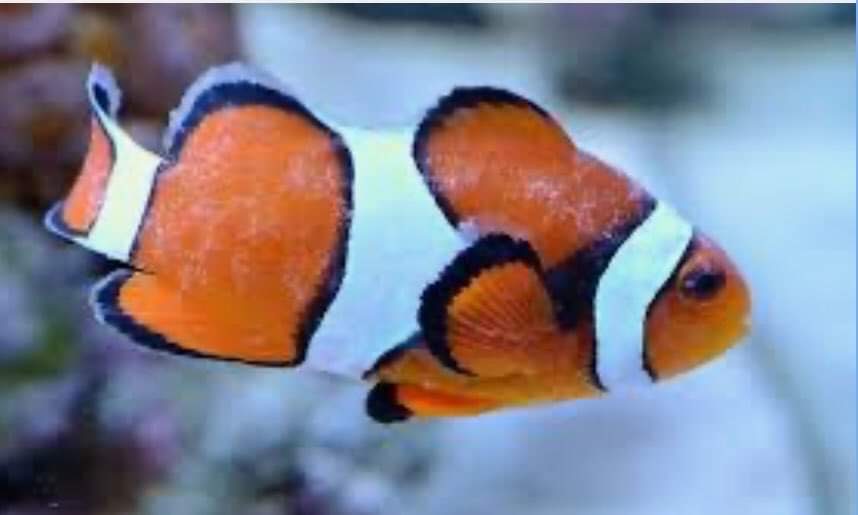
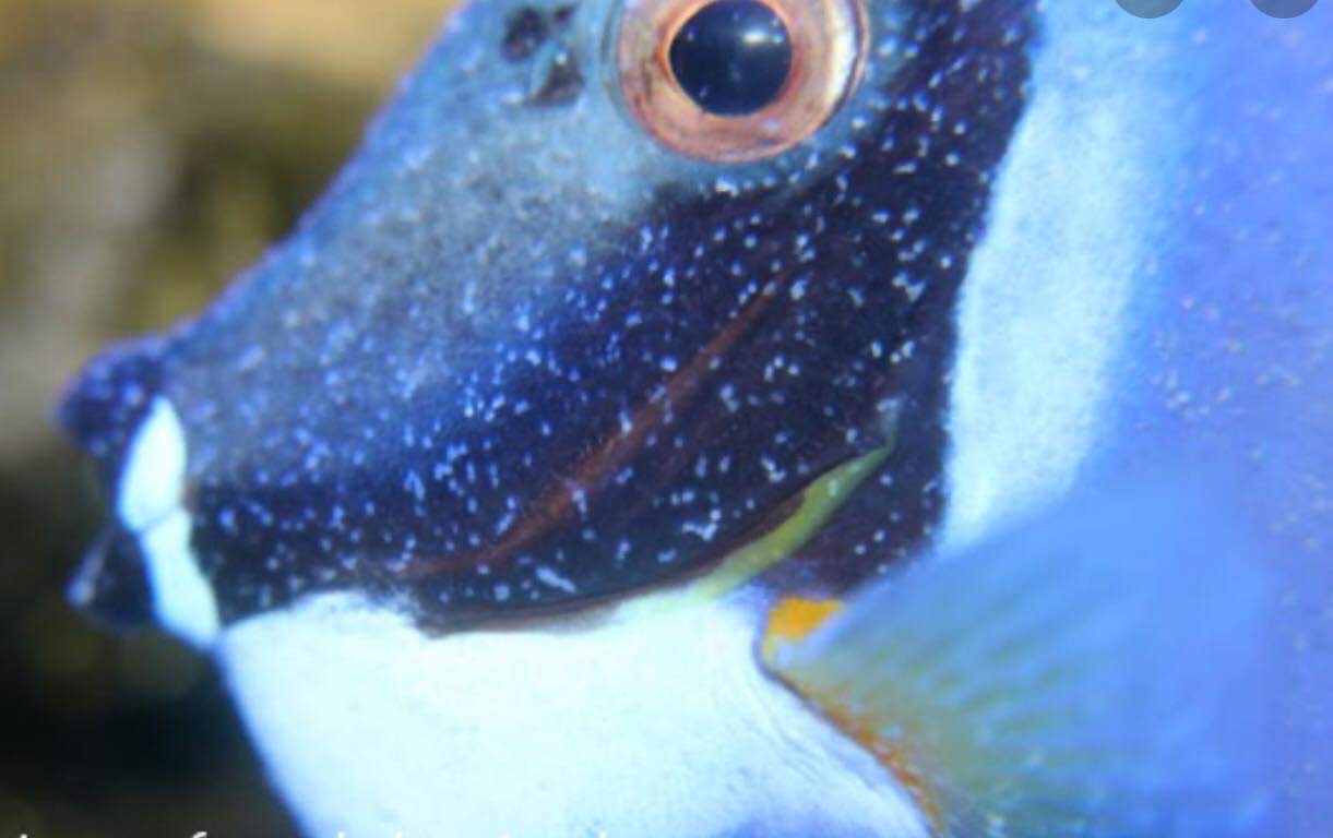
Infected fish therefore appear covered in small white dots, and in the most serious cases, given their high density, they look covered in a sort of white, opaque dust, especially when seen against the light.
Gill breathing is also faster, and affected fish tend to hide.
A fish affected by this parasite can die within a few days if the host is unable to fight off the infection.
How to recognise an affected fish
- 1White dots small, scattered over the body
- 2White, opaque dust in serious cases, clearer against the light
- 3Faster breathing the gills work faster
- 4Hides the fish tends to take shelter
The denser the parasites, the more the dots look like dust: death can come within a few days.
Remedies
As for remedies or cures, it should be stressed that the only method that over the years has shown reasonable chances of success involves the use of copper sulphate, to be used only in a separate tank and under the guidance of a veterinarian.
Since copper sulphate is very toxic to corals and invertebrates, a dedicated quarantine tank must be set up in which to treat the fish.
Copper stays out of the reef
- 1Reef tank corals and invertebrates stay here
- 2No copper here it is very toxic to them
- 3Quarantine tank only the fish are treated here
- 4Copper sulphate used only in the separate tank
The fish move, the copper never goes where corals and invertebrates live.
Sources: [4]
Other remedies, which however do not always give the best results, involve the use of acriflavine or a bath in hypotonic solutions (salinity 1001 ppm).
Recently, the experiences of several aquarists would suggest that the drug fluconazole, an antifungal recently used to rid aquariums of Derbesia and Bryopsis algae, is effective in fighting Oodinium ocellatum infections.
If this evidence were confirmed it would be a great discovery for the hobby, since the drug is well tolerated by corals and invertebrates and using it does not require setting up dedicated quarantine tanks.
What is known about each
Copper sulphate
- For the author: reasonable chance of success
- Toxic to corals and invertebrates
- Doesn't kill the cyst on the bottom
Acriflavine
- For the author: results not always the best
- Mentioned in a study, but not tested
Hypotonic bath
- For the author: results not always the best
- Fresh water reduces parasites on gills and skin
Fluconazole
- Only aquarists' experiences
- No peer-reviewed study found
- To be confirmed
No doses here: choosing a treatment is up to the veterinarian.
Giuseppe Ossolengo
Sources
- WoRMS Editorial Board (2026). Amyloodinium ocellatum (E.-M.Brown) E.-M.Brown & Hovasse, 1946. World Register of Marine Species.
- Smith S.A., Schwarz M.H. (2019). Getting Acquainted with Amyloodinium ocellatum. Virginia Cooperative Extension, Virginia Tech.
- Dhayanithi N.B., Sudhagar A., Ajith Kumar T.T., Lal K.K. (2022). Study on amyloodiniosis outbreak in captive-bred percula clownfish (Amphiprion percula) and improved control regimens. Journal of Parasitic Diseases 46(4): 1103-1109.
- Tedesco P., Beraldo P., Massimo M., Fioravanti M.L., Volpatti D., Dirks R., Galuppi R. (2020). Comparative Therapeutic Effects of Natural Compounds Against Saprolegnia spp. (Oomycota) and Amyloodinium ocellatum (Dinophyceae). Frontiers in Veterinary Science 7.
- Sudhagar A., Sundar Raj N., Mohandas S.P., Serin S., Sibi K.K., Sanil N.K., Swaminathan T.R. (2022). Outbreak of Parasitic Dinoflagellate Piscinoodinium sp. Infection in an Endangered Fish from India: Arulius Barb (Dawkinsia arulius). Pathogens 11(11): 1350.
- Lu Y., Jiang J., Zhao H., Han X., Xiang Y., Zhou W. (2020). Clade-Specific Sterol Metabolites in Dinoflagellate Endosymbionts Are Associated with Coral Bleaching in Response to Environmental Cues. mSystems 5(5): e00765-20.
- U.S. National Library of Medicine (2018). Fluconazole: MedlinePlus Drug Information. MedlinePlus.
On the same topic
Euphyllia: parasites, treatment, husbandry
A garden of Euphyllia is very rewarding, but these corals suffer from swings and parasites. The values to keep, how…
Fluconazole and Oodiniasis
English version of the article Fluconazolo e Oodiniasi: the reports of three reefkeepers, the ergosterol hypothesis…
Fluconazole and marine velvet (Amyloodinium)
Some second-hand experiences suggest that fluconazole may fight marine velvet. The parasite, the ergosterol…